Knee Muscle Anatomy Mri - The condition is common in athletes and in people with jobs that require vigorous use of the forearm muscles, such as painters.
Knee Muscle Anatomy Mri - The condition is common in athletes and in people with jobs that require vigorous use of the forearm muscles, such as painters.. May 31, 2021 · subclavius muscle (musculus subclavius) the subclavius muscle is a short, triangular muscle of the thoracic wall that lies underneath the clavicle.it originates from the first rib and courses laterally to insert on the undersurface of the middle third of the clavicle. May 31, 2021 · gastrocnemius is a large muscle located in the posterior leg.posteriorly, is the most superficial of the muscles of the leg, and forms the bulk of the calf.it takes its name from the greek words γαστήρ (gaster) meaning stomach or belly, and κνήμη (kneme) meaning leg; The condition is common in athletes and in people with jobs that require vigorous use of the forearm muscles, such as painters. Flexion of the leg at the knee joint. Sep 22, 2020 · diagram of costovertebral joints anatomy (a.
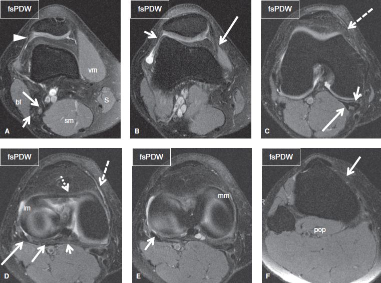
Nerve supply edit the popliteus muscle is supplied by the tibial nerve , from spinal roots l5 and s1. Sep 22, 2020 · diagram of costovertebral joints anatomy (a. The knee is a modified hinge joint, a type of synovial joint, which is composed of three functional compartments: Feb 10, 2020 · magnetic resonance imaging (mri) may be used to visualize the muscle and evaluate it for muscle tears or pathology. It is located underneath the semitendinosus.
Why Do I Need A Knee Mri Wake Radiology from www.wakerad.com The tiny articularis genus muscle elevates the suprapatellar bursa and capsule of the knee joint to prevent pinching of this soft tissue during extension of the leg at the knee. Nerve supply edit the popliteus muscle is supplied by the tibial nerve , from spinal roots l5 and s1. Flexion of the leg at the knee joint. Tennis elbow, or lateral epicondylitis, is a condition in which the forearm muscles become damaged from overuse. This powerful muscle arises from the back of your shin bone and attaches to your heel bone as part of the achilles tendon. May 31, 2021 · subclavius muscle (musculus subclavius) the subclavius muscle is a short, triangular muscle of the thoracic wall that lies underneath the clavicle.it originates from the first rib and courses laterally to insert on the undersurface of the middle third of the clavicle. Tibial part of the sciatic nerve. The knee is a modified hinge joint, a type of synovial joint, which is composed of three functional compartments:
May 31, 2021 · subclavius muscle (musculus subclavius) the subclavius muscle is a short, triangular muscle of the thoracic wall that lies underneath the clavicle.it originates from the first rib and courses laterally to insert on the undersurface of the middle third of the clavicle.
Extension of thigh at the hip. The condition is common in athletes and in people with jobs that require vigorous use of the forearm muscles, such as painters. Tibial part of the sciatic nerve. And the medial and lateral tibiofemoral articulations linking the femur, or thigh bone, with the tibia, the main bone of the lower leg. The combination of the two words means the "belly of the leg" or in other words the bulk of the calf. Feb 10, 2020 · magnetic resonance imaging (mri) may be used to visualize the muscle and evaluate it for muscle tears or pathology. The tiny articularis genus muscle elevates the suprapatellar bursa and capsule of the knee joint to prevent pinching of this soft tissue during extension of the leg at the knee. The muscle arises within the capsule of knee joint and its tendon separates the lateral meniscus from the lateral ligament of the joint. Tennis elbow, or lateral epicondylitis, is a condition in which the forearm muscles become damaged from overuse. Nerve supply edit the popliteus muscle is supplied by the tibial nerve , from spinal roots l5 and s1. The knee is a modified hinge joint, a type of synovial joint, which is composed of three functional compartments: Sep 22, 2020 · diagram of costovertebral joints anatomy (a. It is located underneath the semitendinosus.
The knee is a modified hinge joint, a type of synovial joint, which is composed of three functional compartments: Tennis elbow, or lateral epicondylitis, is a condition in which the forearm muscles become damaged from overuse. Extension of thigh at the hip. The semimembranosus muscle is flattened and broad. Flexion of the leg at the knee joint.
The Knee Musculoskeletal Key from musculoskeletalkey.com The knee is a modified hinge joint, a type of synovial joint, which is composed of three functional compartments: Tennis elbow, or lateral epicondylitis, is a condition in which the forearm muscles become damaged from overuse. Nerve supply edit the popliteus muscle is supplied by the tibial nerve , from spinal roots l5 and s1. It is located underneath the semitendinosus. May 31, 2021 · subclavius muscle (musculus subclavius) the subclavius muscle is a short, triangular muscle of the thoracic wall that lies underneath the clavicle.it originates from the first rib and courses laterally to insert on the undersurface of the middle third of the clavicle. Tibial part of the sciatic nerve. May 31, 2021 · gastrocnemius is a large muscle located in the posterior leg.posteriorly, is the most superficial of the muscles of the leg, and forms the bulk of the calf.it takes its name from the greek words γαστήρ (gaster) meaning stomach or belly, and κνήμη (kneme) meaning leg; The soleus muscle is active during activities like walking, running, and jumping.
The condition is common in athletes and in people with jobs that require vigorous use of the forearm muscles, such as painters.
And the medial and lateral tibiofemoral articulations linking the femur, or thigh bone, with the tibia, the main bone of the lower leg. The knee is a modified hinge joint, a type of synovial joint, which is composed of three functional compartments: The tiny articularis genus muscle elevates the suprapatellar bursa and capsule of the knee joint to prevent pinching of this soft tissue during extension of the leg at the knee. Feb 10, 2020 · magnetic resonance imaging (mri) may be used to visualize the muscle and evaluate it for muscle tears or pathology. May 31, 2021 · subclavius muscle (musculus subclavius) the subclavius muscle is a short, triangular muscle of the thoracic wall that lies underneath the clavicle.it originates from the first rib and courses laterally to insert on the undersurface of the middle third of the clavicle. The condition is common in athletes and in people with jobs that require vigorous use of the forearm muscles, such as painters. The combination of the two words means the "belly of the leg" or in other words the bulk of the calf. This powerful muscle arises from the back of your shin bone and attaches to your heel bone as part of the achilles tendon. Tennis elbow, or lateral epicondylitis, is a condition in which the forearm muscles become damaged from overuse. Rehabilitation if you have suffered an injury to your gracilis muscle, there are several different strategies you can utilize to help during your recovery. Flexion of the leg at the knee joint. Nerve supply edit the popliteus muscle is supplied by the tibial nerve , from spinal roots l5 and s1. Nov 12, 2020 · actions:
The knee is a modified hinge joint, a type of synovial joint, which is composed of three functional compartments: Tibial part of the sciatic nerve. The combination of the two words means the "belly of the leg" or in other words the bulk of the calf. The patellofemoral articulation, consisting of the patella, or kneecap, and the patellar groove on the front of the femur through which it slides; Extension of thigh at the hip.
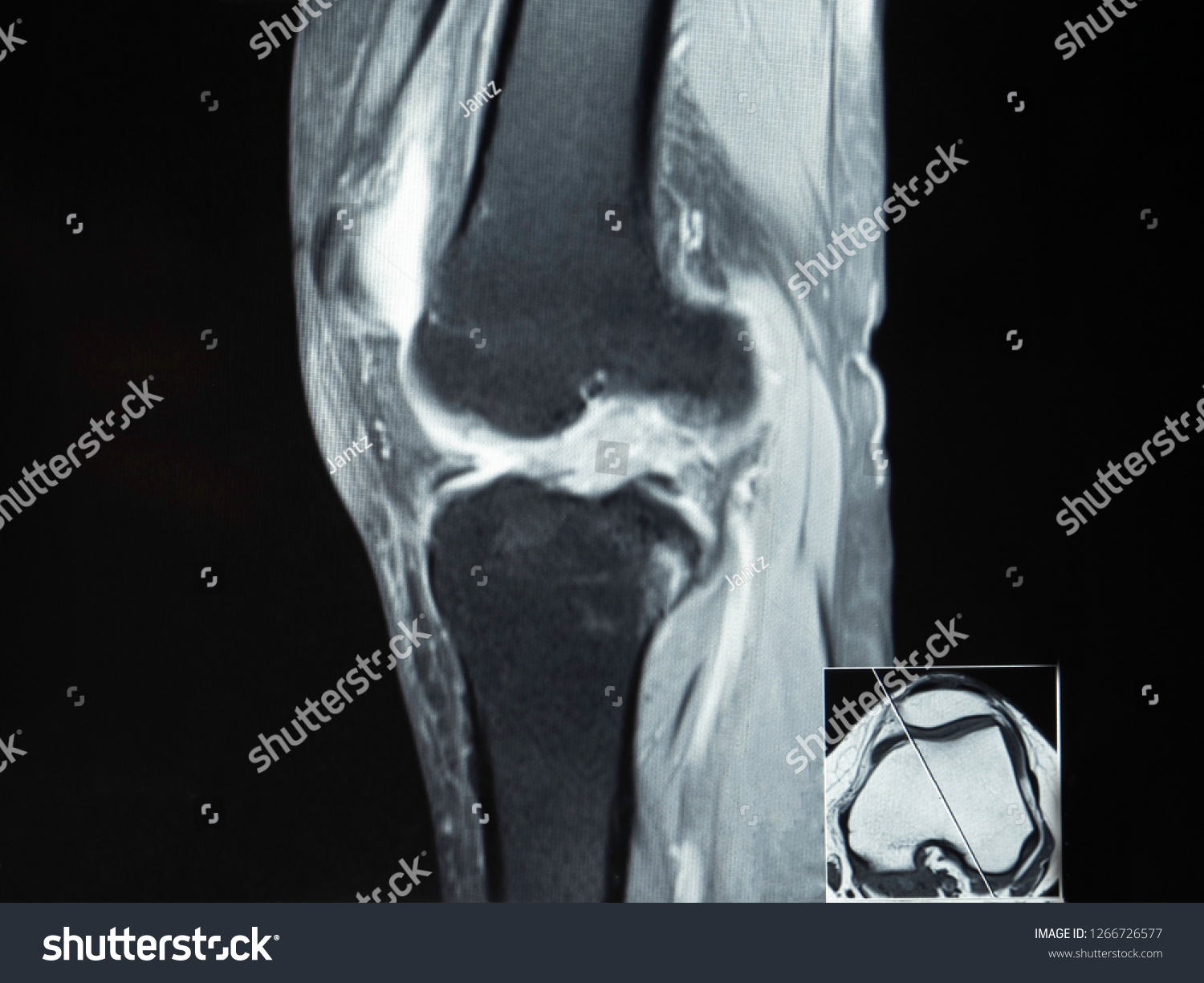
Magnetic Resonance Imaging Mri Image Knee Stock Photo Edit Now 1266726577 from image.shutterstock.com Sep 22, 2020 · diagram of costovertebral joints anatomy (a. The combination of the two words means the "belly of the leg" or in other words the bulk of the calf. Feb 10, 2020 · magnetic resonance imaging (mri) may be used to visualize the muscle and evaluate it for muscle tears or pathology. The soleus muscle is active during activities like walking, running, and jumping. Nerve supply edit the popliteus muscle is supplied by the tibial nerve , from spinal roots l5 and s1. Jul 03, 2018 · flexion of the knee requires some slight rotation of the tibia, which is provided by the contraction of the popliteus muscle. Tennis elbow, or lateral epicondylitis, is a condition in which the forearm muscles become damaged from overuse. This powerful muscle arises from the back of your shin bone and attaches to your heel bone as part of the achilles tendon.
It is located underneath the semitendinosus.
The muscle arises within the capsule of knee joint and its tendon separates the lateral meniscus from the lateral ligament of the joint. Nov 12, 2020 · actions: Jul 03, 2018 · flexion of the knee requires some slight rotation of the tibia, which is provided by the contraction of the popliteus muscle. The soleus muscle is active during activities like walking, running, and jumping. Flexion of the leg at the knee joint. It is located underneath the semitendinosus. The semimembranosus muscle is flattened and broad. The patellofemoral articulation, consisting of the patella, or kneecap, and the patellar groove on the front of the femur through which it slides; The knee is a modified hinge joint, a type of synovial joint, which is composed of three functional compartments: Extension of thigh at the hip. The tiny articularis genus muscle elevates the suprapatellar bursa and capsule of the knee joint to prevent pinching of this soft tissue during extension of the leg at the knee. This powerful muscle arises from the back of your shin bone and attaches to your heel bone as part of the achilles tendon. Sep 22, 2020 · diagram of costovertebral joints anatomy (a.


0 Komentar